Why does space
between top front teeth keep opening up after orthodontic treatment?
Probably one of the most noticeable
malocclusions in people with otherwise straight teeth is that of spacing
between upper incisors. In fact, there
usually is fairly good alignment of the upper teeth in these cases so many
times patients and parents think this is an easy fix. Many may even try to close space with an
upper clear aligner or upper braces alone only to find the space(s) return soon
after closure.
Space in the upper front teeth as an
adolescent (after the upper cuspids have erupted) when there is no space in the
lower can be a sign of heavy occlusion of the front teeth which in turn holds
or pushed the upper front teeth outward; as the arch circumference increases
(but the tooth sizes remain constant), the patient experiences gaps, usually
between upper central incisors and lateral incisors. It can be gradual or even develop as the
patient transitions from baby teeth to permanent teeth. If the patient is waiting on upper canines to
erupt, the space may close when these canines fully erupt. If the space is present after canines erupt,
then there is usually a problem. Over
time, heavy contact can lead to excessive wear of the teeth, mobility or even
recession.
This heavy contact can be from
excessive growth of the lower jaw forward (which brings lower teeth forward
essentially “jamming” them into the back of upper incisors), upper front teeth
leaning backward (termed “Division II”) or can from smaller than average upper
teeth. In rare occasions there can be an
extra tooth under the gums which must be removed. The sequence of eruptions and the timing of
the lower jaw growth can also lead to a deepbite (early over-eruption of lower
incisors) which also can cause heavy contact and spacing.
 |
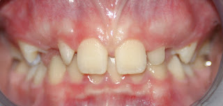
| Deepbite with premature contact on upper incisors. This patient also presents with congenitally small lateral incisors making the space even larger between from teeth. |
 |
|
Following
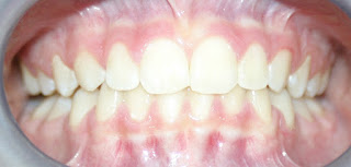
treatment with braces, spaces between front teeth are closed, the deepbite is
corrected and the small lateral incisors have been built up with white “composite”
material.
|
Sometimes the upper front teeth grow
in and lean away from each other making the appearance of a large gap (see
patient below); there may be contact but is hidden under the gumline. In these cases, the teeth must be uprighted
so that they lean back toward each other, this brings the contact point from
below the neckline (below the gums) up to the top 1/3 of each tooth which can
completely close the space. This is
something I see left in cases after treatment as patients come in with
retainers “that won’t close the space” from other offices. I have to explain to these patients that the
teeth ARE in contact but it is below the gumline; to “close” the visible space we
have to attach brackets to the teeth and tip them back toward each other as we
shift the roots away from each other. No
retainer can close space when the teeth are leaning away from each other and
already in contact below the gumline.
So how can
we fix the spacing and keep it closed?
If upper incisors are leaning backward
then these upper front teeth must be uprighted first; this is a different type
of case and is covered in other articles due to the unique complications of
that malocclusion.
 |
|
Same
patient after correcting front teeth, notice the space closes easily once the
occlusion allows the teeth to bite normally.
|
 |
|
Cephalometric
Radiograph to asses tipping of front teeth (Division II)
|
But if the front teeth are not leaning
backward (Orthodontists use a cephalometric or side-view X-Ray to determine and
quantify this angle) then the answer is we must focus on the lower teeth
first. In almost EVERY case of heavy incisor
contact and spacing in the upper arch, the first step is to pull lower front
teeth back, away from the upper front teeth.
Once pulled back and there is room for upper teeth to also be brought
back, then the upper space between incisors can be closed and held. Patients should be given a couple of months at
the end (before removing appliances) to allow the lower jaw to shift or “settle”
because it may have been pushed back and held back by the patient subconsciously
so that once it gains the freedom to move, it may come forward bringing the
lower teeth forward back into contact; this requires more retraction of the
lower teeth or the space will return.
If lower jaw growth is significant
enough, and the patient young enough, we may consider bringing the upper jaw
forward (see my article on when to treat underbites).
 |
|
This
7 year old patient presented with an early developing underbite; correction
required pulling the entire upper jaw forward with protraction headgear (see
blog on when to fix underbites).
|
 |
|
At 12
years old, following correction of the underbite from age 7 to 8, the patient
no longer has space or heavy contact on front teeth. In fact, no further treatment was needed or recommended.
|
Growth later in life can increase
space that used to be minimal; the patient below is an example of exceptional
lower jaw growth that was not corrected early as in the above patient.
Finally, once the lower teeth are
pulled back (and aligned) and upper space closed and held to allow time for the
lower jaw to shift, then retainers must be placed and monitored closely for the
next 6 to 12mo to check for any heavy contact returning on individual
teeth. If a single tooth is still mobile
3mo following braces, you can safely assume heavy contact and relapse of space;
this tooth should be marked with articulating paper (typewriter ribbon for us
older folks) and adjusted with judicial polishing of the enamel in the
offending spot(s).
If the patient can tap/bite on their
back teeth and feel no movement in the upper front teeth, they are safe to go
back to regular retainer wear. But this
should be checked regularly over the first year (and even further depending on
the patient’s age and growth pattern) if you expect to keep the spaces closed.
“I have
heard that the gums between front teeth must be cut to close the space,” what
about this?
In the past, cutting the gum tissue
between teeth with large space in the midline (frenectomy) was sometimes
performed if the attachment of the lip was high and between the teeth (similar
to being tongue-tied in the lower arch).
When this attachment is the problem, it is very deep and will require an
oral surgeon to separate the tissue all the way to the bone (See picture below). Unfortunately this is almost never done completely
during “typical” frenectomies. In truth,
the tissue between two front teeth that have a gap is usually just filling the
space because the space is already there; it is not normally the cause of the
space.
 |
|
These
pictures illustrate a before and after frenectomy due to attachment of the
frenum directly between the front central incisors.
|
Does that
mean patients never need a frenectomy?
No. Sometimes the gap has been present
so long that when the teeth are brought together, the tissue “bunches up” and
becomes chronically inflamed. In these
cases, the excess soft tissue can be removed following space closure using a
laser with minimal discomfort and quick healing.
My doctor
wants everyone to have a laser frenectomy, is that wrong?
 |
|
A
good case from relieving the tissue that connect the lip to the gums (frenum or
frenulum); if left to during development, this thick tissue will hold front
teeth apart.
|
I believe there is a trend in offices
that get lasers to be more aggressive in prescribing laser frenectomies. There is some logic in reducing this tissue
in some cases (see above) but until lasers, we just rarely sent out
frenectomies for anything less than the most significant space. I have seen no evidence that routine laser
frenectomies are effective in most cases of spacing but I will continue to
monitor the journals for a juried study to come out. I will say that the sales people that market
these soft-tissue lasers certainly stress the profitability based on X
percentage of patients getting laser frenectomies every week which makes me a
little wary if not somewhat nauseous.
 |
|
This photo
(borrowed from the Internet) shows a “successful” frenectomy that has healed
nicely but you can see there is no effect on the initial spacing from removal
of the frenum.
|
I do like the lasers for more
surface-based frenectomies, they are faster than the scalpel and have less side
effects/bleeding/discomfort, but I have seen early recession in one case so for
now and as I mentioned previously, true cases of frenum attachment goes to the
bone which requires actual surgery and not just a surface release of soft
tissue. So I am personally reserving
referrals of my patients to really obvious and significant cases only. And I rarely refer until the space is closed
to prevent scar tissue from building which may actually make space closure more
difficult.


Thanks for taking the time to share this wonderful information with us. I enjoyed the details that you provided. Have a great rest of your day.
ReplyDeleteDentist Philadelphia
This blog was extremely useful. I really appreciate your kindness in sharing this with me and everyone else! upper teeth.
ReplyDelete